Surgical Instructions
Even though most dental surgeries are performed as outpatient procedures, they require specific preparation and care afterward. To ensure that you receive safe and comfortable care, we ask that you take the time to read our pre- and post-operative instructions, which are intended to help you have a smooth operation and a faster recovery.
We would be glad to answer any questions or concerns you may have regarding your surgery by contacting our practice as soon as possible.

During a routine oral cancer examination, the following signs will be examined:
Red patches and sores
The presence of red patches on the floor of the mouth, the front and sides of the tongue, as well as white or pink patches that do not heal and sores that heal slowly and bleed easily may be indicative of pathologic (cancerous) changes.
Leukoplakia
This is a hardened, white or gray, slightly raised lesion that can occur anywhere in the mouth. In the absence of treatment, leukoplakia may develop into cancer.
Lumps
The presence of soreness, lumps, or thickening of tissues anywhere in the throat or mouth can indicate a pathological condition.
Oral cancer - Exams, Diagnosis and Treatment
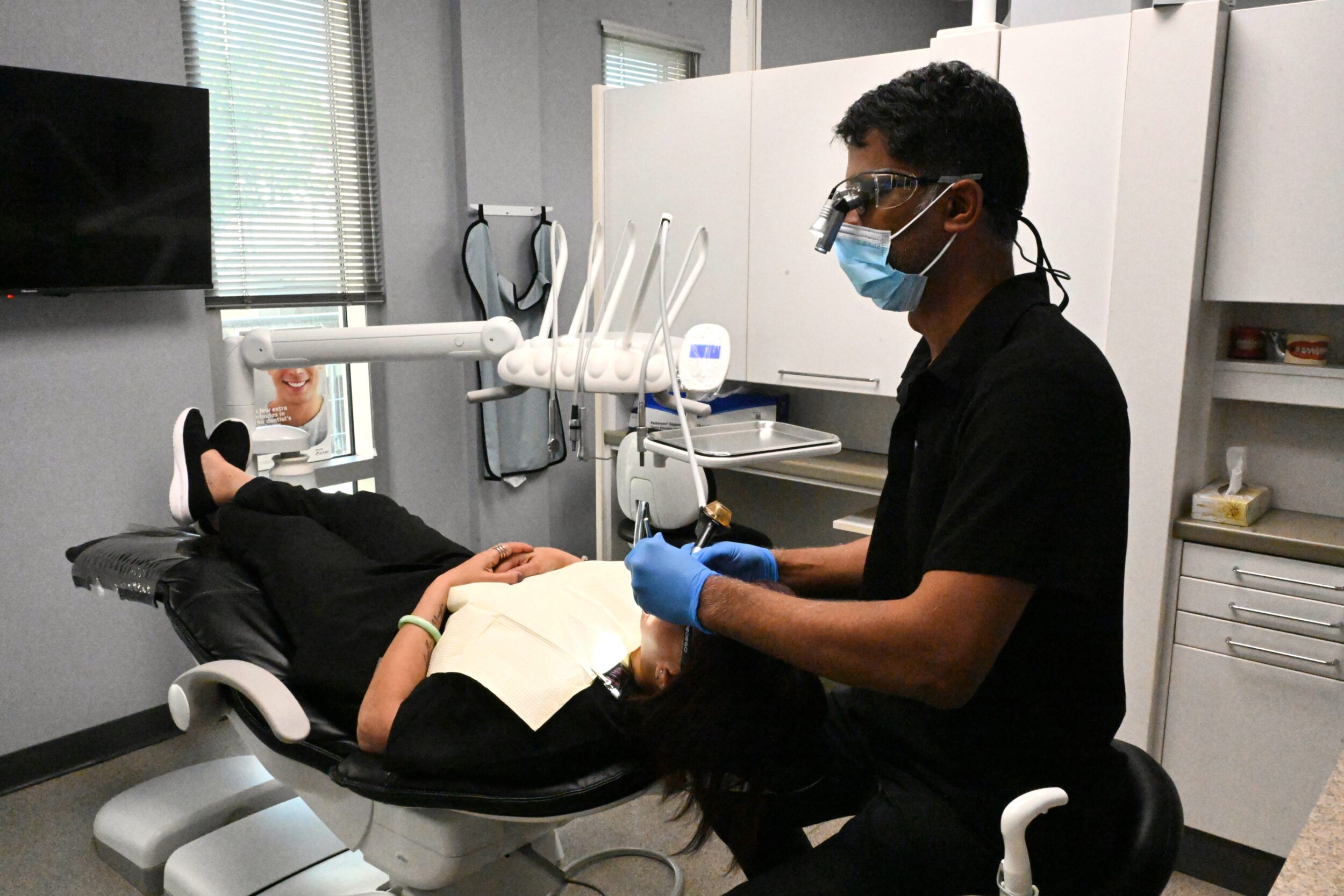
Oral cancer examinations are completely painless. It is during the visual part of the examination that the dentist will look for abnormalities, as well as feel the face, glands and neck to look for unusual bumps. A laser that can highlight pathologic changes in the oral cavity is also a wonderful tool for the detection of oral cancer. A laser can be used to detect abnormal signs and lesions below the surface that are not visible to the naked eye.
A diagnostic impression and treatment plan will be implemented if abnormalities, lesions, leukoplakia or lumps are evident. It is possible that a biopsy of the area will be needed in the event that the initial treatment plan does not work. In addition to the biopsy, a clinical evaluation will determine the exact stage and grade of the oral lesion.
When the basement membrane of the epithelium has been ruptured, oral cancer is considered to be present. Cancers of the oral and maxillofacial regions can readily spread to other parts of the body, posing additional secondary threats. It is important to note that treatment methods may vary depending on the specific diagnosis, but may include excision, radiation therapy, and chemotherapy.
As part of the dentist and hygienist’s bi-annual checkup, the dentist and hygienist will thoroughly check for changes and lesions in the mouth, but an oral cancer screening should be conducted at least once a year to detect any abnormalities.
Ask your dentist or dental hygienist if you have any questions or concerns about oral cancer.